
Teaching Microscopy Using Plants

This series of articles from Dr Chris Graham (Visiting Teacher at SAPS) explore how plant specimens can be used to support teaching microscopy.
The first article, What does a microscope show me?, explores the basics of how a microscope works, building student confidence.
The second article, Preparing specimens, which can be found further down this page, demonstrates why specimens must be thin and the role of staining, better preparing students for making their own slides.
Article 1 – What does a microscope show me?
The first article, published in the November 2024 issue of School Science Review, suggests readily available plant material that can be used to help students of any age become confident and enthusiastic users of microscopes as well as skilled interpreters of what they see. The specimens discussed require minimal preparation and no staining, allowing students to spend more time using microscopes and less time preparing slides. What is viewed under the microscope is also easily relatable to what students can see with the naked eye, thereby minimising the conceptual leap needed to interpret what is being viewed under the microscope.
The article explores:
- the concepts of depth of field, focal plane, and field of view
- how the leaves of Tradescantia zebrina (silver inch plant) and the pondweed Egeria densa can be used to build students’ understanding of these concepts
- how using a dandelion seed (Taraxacum sp.) can show that a microscope creates an optical slice through a specimen.
These specimens also help students gain a sense of scale while using the microscope to transition from the ‘just visible with the naked eye’ to ‘only visible with a microscope’.
The associated PowerPoint presentation contains high resolution images you can use in your teaching.
You can also download our microscopy troubleshooting guide for teachers and technicians to help you support your students.
The protocols for each of the techniques can be found on the SAPS website:
Microscopy of a dandelion seed: exploring depth of field and focal plane
Observing stomata in Tradescantia zebrina
Microscopy of a pondweed leaf: exploring depth of field and focal plane
The videos below demonstrate two of the specimens suggested in the first article, What does a microscope show me?
Cytoplasmic streaming in pondweed (Egeria densa)
You must accept YouTube marketing cookies before seeing this video.
This video starts by focusing though the full depth of a leaf of the pondweed Egeria densa. The leaf is two cells thick and the first part of the video focuses from the top of the leaf to the bottom and back up again to allow the viewer to build up an understanding of the 3D structure of the leaf. (Note: these sections have been sped up slightly.)
The video then shows the movement of chloroplasts at two different depths within the leaf (in real time).
Firstly, chloroplasts are seen moving across the uppermost layer of cytoplasm in the lower layer of cells, and secondly chloroplasts are seen streaming around the edge of the cells in the thin layer of cytoplasm between the plasma membrane and the vacuole. This cytoplasmic streaming is thought to help these large cells overcome the limitations of diffusion by constantly stirring the contents of the cell. It also shows that cells are dynamic, living things and not just static objects.
Exploring focal plane with a dandelion seed
You must accept YouTube marketing cookies before seeing this video.
This video was created by focussing through the pappus (the filamentous parachute-like structure) of a dandelion seed (Taraxacum sp.).
It was filmed by securing a dandelion seed horizontally in some Blu Tack on a slide without a coverslip. You can see this setup in the first part of the video.
It shows that only part of a specimen is in focus at any one time and that objects just out of focus are noticeable but blurry whereas objects that are far away from the focal plane are not noticeable at all.
You can find the protocol for this activity here.
Article 2 – Preparing specimens

The second article, published in the March 2025 issue of School Science Review, continues the discussion about supporting the development of students’ microscopy techniques by exploring the two most fundamental components of slide preparation that allow the investigation of a much wider range of biological material: the importance of a thin specimen and the value of achieving contrast through staining. With a secure appreciation of slide preparation, students should be able to produce better preparations of stem sections and root tip squashes and be able to focus more on the biology that they are exploring rather than the methods of slide preparation.
The article explores:
- the importance of obtaining a thin specimen through observation of red bell pepper
- the use of staining to provide contrast when investigating starch content in smears from ripe and unripe bananas
- the value of staining for identification of molecular make-up when investigating sclereid cells in pear
The associated PowerPoint presentation contains high resolution images you can use in your teaching.
The protocols for each of the techniques can be found on the SAPS website:
Microscopy of red bell pepper skin: the importance of having a thin specimen
Microscopy of a banana smear: using a stain to provide contrast
Microscopy of pear pulp sclereids: staining to identify molecules
Article 3 – Calibrating and measuring
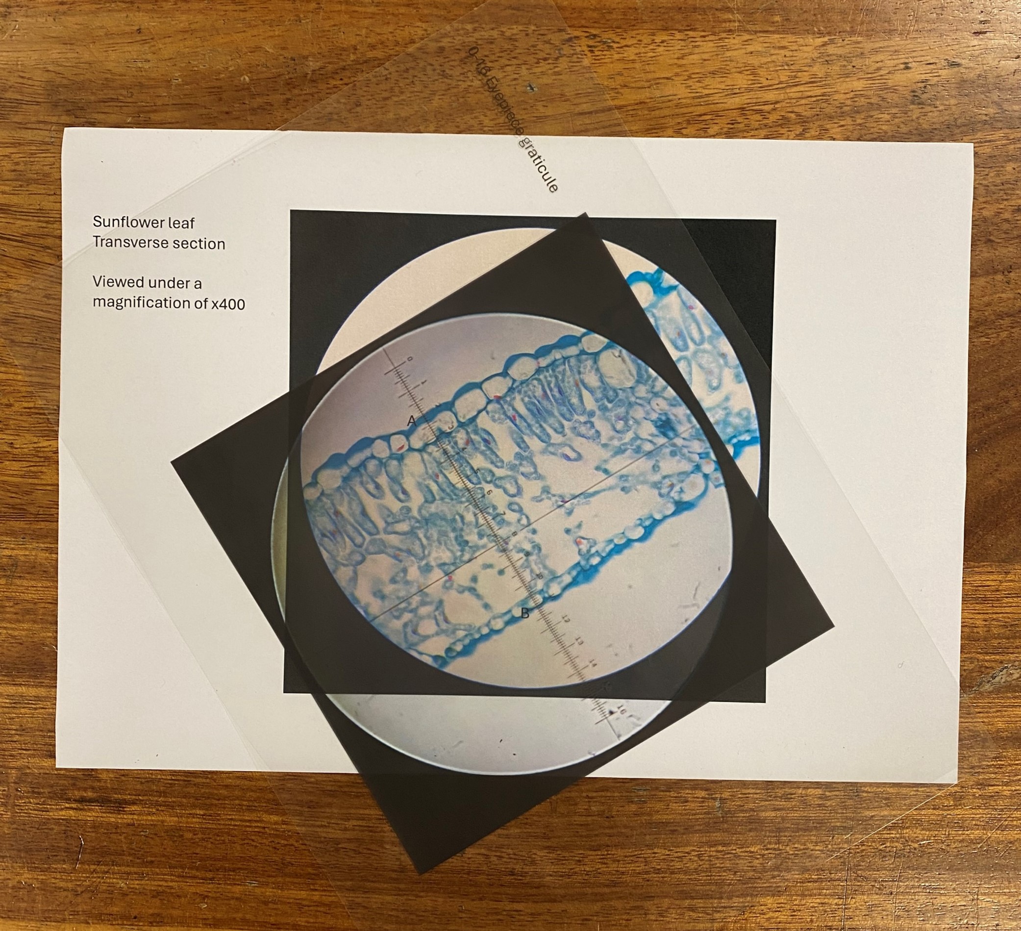
The third article, published in the July 2025 issue of School science Review, focuses on skills that tend to be required of older students. However, with minor modifications, most of the preparations discussed in this article could be used with younger students as well. This article suggests four activities to support the development of calibrating and measuring skills. To allow as much time as possible for developing these skills, the specimens suggested here require minimal preparation.
The article begins with a paper-based activity that supports students in learning to use an eyepiece graticule and a stage micrometer before going on to describe three investigations that students can carry out whilst developing these skills using a microscope.
Students can then use their measuring and calibrating skills to investigate:
- What happens to cells as an onion grows?
- What happens to stomatal density as a leaf grows?
- How fast are the chloroplasts moving inside pondweed cells?
The protocols for each of the techniques can be found on the SAPS website:
Calibrating and Measuring in Microscopy – a paper-based activity
Exploring Growth in Onion Epidermis
Receive the latest resources and updates
Get half-termly email newsletters with new resources, CPD opportunities, plant science news and inspiration.
Sign up now

